

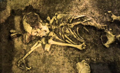
For many years, archaeologists have encountered a puzzling occurrence: skeletons degraded over time where the only remaining soft tissue is surprisingly the brain. Recently, a group of scientists has revealed the chemical processes responsible for this extraordinary preservation. Their findings suggest that it’s not a rare preservation fluke but rather a consequence of how decay unfolds.
This enigma, referred to as the brain preservation paradox, has intrigued researchers for decades. Research led by Alexandra Seviour, a paleobiology PhD candidate at the University of Oxford, has documented over 4,400 well-preserved human brains spanning 12,000 years in prehistoric contexts. The ability of brain tissue to endure while other tissues like skin and muscle vanish has defied typical understanding of decomposition.
What makes this discovery even more surprising is the typical environments in which these preserved brains are found. Roughly one-third have been recovered from saturated, low-oxygen settings such as riverbeds, lakesides, submerged caves, and shipwrecks — areas usually associated with rapid decay rather than conservation.

Six-Month Burial Experiment Yields Insights
In a novel experiment published on June 19 in the Journal of Proteome Research, researchers buried mouse bodies under varying combinations of moisture and oxygen availability, then tracked the degradation over a half-year period.
Examining the brain tissues at intervals — including 24 hours, 72 hours, 1 week, 6 weeks, 3 months, and 6 months — the team employed advanced mass spectrometry techniques to pinpoint which proteins remained stable and which deteriorated.
“We were looking for which specific peptides survived and which vanished, and what chemical marks were left on the survivors,” Alexandra Seviour explained.

From their extensive data set of over 1.26 million patterns of protein degradation, the researchers observed similar decay rates across initial burial conditions. However, differences in oxygen availability became apparent after several weeks.
Higher oxygen levels accelerated brain tissue decay, whereas damp, oxygen-deprived environments facilitated the formation of robust protein configurations that resist breakdown. These protective structures could account for the survival of ancient brain tissue over millennia.
The Role of Free Radicals in Preservation
Seviour pointed out that free radicals — highly reactive molecules that damage proteins and cells — play a crucial role. In oxygen-rich conditions, these molecules promote rapid protein destruction and tissue decay. Conversely, in wet, low-oxygen settings, the breakdown slows considerably.
Rather than fully degrading proteins, some chemical byproducts adhere to them, forming stable and resilient molecular frameworks that resist further deterioration.

Brains possess features perfectly suited for this kind of preservation: they contain metal ions that facilitate these chemical reactions, fatty membranes conducive to free radical accumulation, and specific amino acids that trap radicals and bolster protective cross-links. The skull structure further aids preservation by limiting oxygen and fluid circulation, compared to other body parts.
Unexpected Connections to Alzheimer’s Research
Richard Evershed, an organic geochemist at the University of Bristol unaffiliated with the study, praised the work’s thoroughness. He noted:
“Comparing more tissues — other organs and muscles — would be really useful to get an idea whether what was happening in the brain was special compared to what was happening elsewhere, and also to resolve questions regarding proteins preserved in other environments in archaeology such as pots or dental calculus.”

Seviour also highlighted that the molecules in these decay-resistant brain proteins resemble those observed in neurodegenerative conditions like Alzheimer’s disease. Further research is needed to explore if these parallels indicate a deeper biological connection.
- Categories:
- Science








0 comments
Sign in to Comment